
DAMAGED TEETH · MIDTOWN MANHATTAN
Do You Have a Cavity or Is Something Else Causing the Tooth Change?
Cavities can be completely painless at first. A white or dark spot, sweet sensitivity, food trapping, a rough area or a visible hole can all deserve attention but color or pain alone cannot tell you whether tooth decay is present or how deeply it extends.
No pain does not rule it out
Early decay can develop before the tooth becomes sensitive.
A dark spot is not a diagnosis
Staining, grooves and arrested changes can resemble active decay.
Book Appointment
Get Your Consultation
Same-day replies. No obligation. A real dentist reviews every request.
Tooth decay and a physical cavity are related, but they are not exactly the same stage. Early mineral loss can exist while the tooth surface is still intact. That distinction matters because selected non-cavitated lesions may sometimes be arrested or remineralized, while tooth structure already lost to a physical cavity does not grow back to its original shape.
20-SECOND EXPLANATION
What Is Tooth Decay and When Does It Become a Cavity?
Tooth decay is a biofilm-mediated disease process in which repeated acid attacks shift the tooth toward mineral loss. A cavity is the structural result when enough hard tooth tissue has been lost that the surface breaks down.
01
Early decay begins as mineral loss
The enamel may look chalky or different without having a physical hole.
02
Saliva and fluoride support repair of early mineral changes
When the surface is still intact, prevention and remineralization strategies may help arrest the process in selected lesions.
03
Cavitation means structure has been lost
Once the tooth surface has physically collapsed, toothpaste cannot rebuild the missing shape.
WHAT ARE YOU NOTICING?
Common Signs of Cavities and Tooth Decay
Some cavities cause no symptoms. Others become noticeable only after decay reaches dentin, affects a restoration or moves closer to the pulp.
A white or chalky spot
An intact white-spot lesion can reflect mineral loss, but not every white area is active decay.
A brown or dark area
Active decay, an inactive/arrested area, stain or a normal dark groove can look similar without a clinical exam.
A visible hole or rough pit
A physical defect can indicate cavitation, broken enamel or a restoration problem and should be evaluated.
Sweets or cold trigger one tooth
Localized sensitivity can occur when decay reaches dentin, although exposed dentin and other conditions can feel similar.
Food keeps getting stuck in one spot
Repeated food trapping can occur with decay between teeth, a damaged contact or a failing restoration.
The tooth now aches or swells
Lingering, spontaneous or swelling-related symptoms can mean the problem has moved beyond an early surface lesion.
HOW DECAY CAN PROGRESS
From Early Mineral Loss to Deeper Tooth Involvement
Not every lesion passes through every stage, and progression speed varies. The purpose of this section is to explain the possible pathway not predict what will happen to a specific tooth.
Healthy / balanced enamel
Mineral loss and repair remain in balance; the tooth surface is intact.
Early demineralization
Minerals are being lost from enamel, sometimes creating a chalky white area without a hole.
Enamel cavitation
The surface loses enough structure that a physical defect or cavity forms.
Decay into dentin
The lesion extends into the softer layer beneath enamel, where sensitivity may become more noticeable.
Pulp / infection involvement
Advanced decay can irritate or infect the inner tooth, sometimes causing lingering pain, spontaneous pain or swelling.
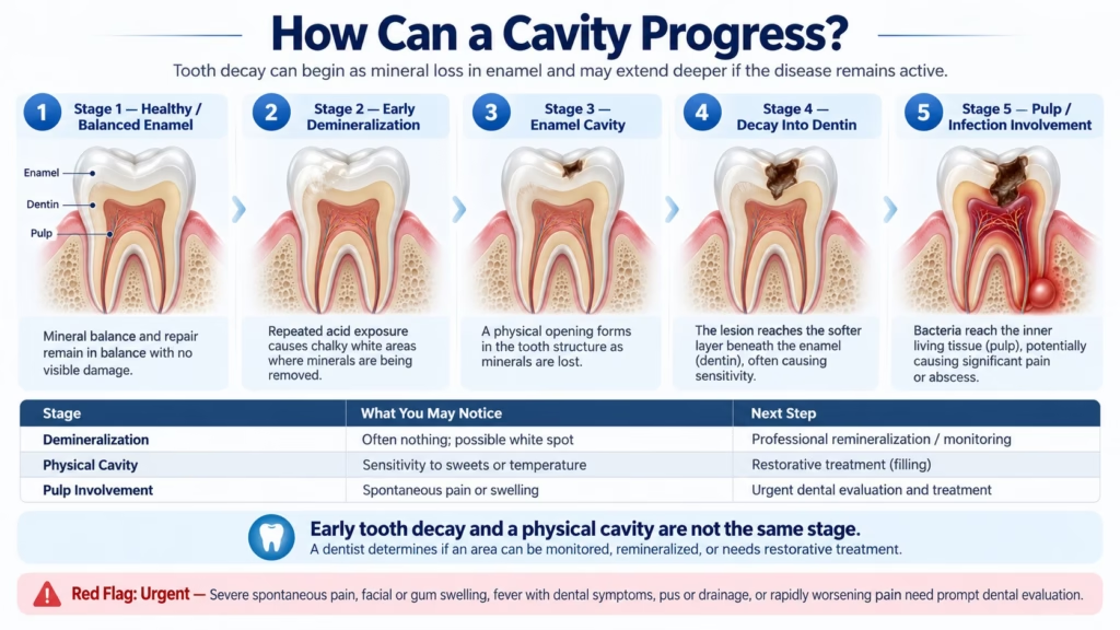
Early Decay and a Physical Cavity Are Not the Same Stage
An intact early lesion may sometimes be managed without drilling. Once a physical hole has formed, the missing tooth shape does not naturally regenerate and the tooth needs professional evaluation to decide how it should be managed.
No pain can still occur
Early lesions and even some cavitated areas may be symptom-free.
Symptoms overlap
Sweet, cold and chewing discomfort can have causes other than decay.
Depth changes the plan
Monitoring, fluoride, filling, larger restoration or pulp treatment are different clinical pathways.
A KEY TREATMENT DISTINCTION
Early Demineralization vs. a Physical Cavity
This is one of the most important ideas on the page because it explains why not every early decay lesion automatically needs a filling.
Early Non-Cavitated Lesion
Minerals have been lost from enamel, but the surface has not collapsed into a hole.
- Often painless.
- May appear white/chalky when active, though appearance alone is not diagnostic.
- Selected lesions may be arrested or remineralized with fluoride, improved plaque control and risk-factor changes.
- Needs professional monitoring because activity and location matter.
Cavitated Lesion / Cavity
The hard tooth surface has physically broken down and a structural defect is present.
- May still be painless, especially before deeper tissues become involved.
- Can trap plaque and food more easily.
- The missing tooth shape does not grow back naturally.
- Professional management is needed to determine whether restorative treatment is appropriate.
WHY IT HAPPENS
What Can Increase the Risk of Tooth Decay?
Cavities are multifactorial. Frequency of carbohydrate exposure, plaque biofilm, saliva, fluoride, tooth anatomy and existing dental work all influence risk.
Repeated acid challenges can outpace the tooth's ability to recover minerals.
Frequent fermentable carbohydrates
Sugary or starchy foods/drinks repeatedly feed acid-producing plaque bacteria.
Saliva has less recovery time
Frequent snacking or sipping can keep the oral environment acidic for longer periods.
Net mineral loss develops
If demineralization repeatedly exceeds repair, the lesion can progress.
Frequent sugar / starch exposure
How often teeth are exposed to fermentable carbohydrates matters because repeated acid attacks reduce recovery time.
Plaque accumulation
Hard-to-clean grooves, crowded contacts and areas around restorations can retain plaque biofilm.
Dry mouth / reduced saliva
Saliva helps neutralize acids and return minerals to enamel. Reduced flow can increase decay risk.
Limited fluoride exposure
Fluoride supports remineralization and helps enamel become more resistant to future acid challenges.
Exposed root surfaces
Recession can expose root surfaces that do not have the same enamel protection as the crown of the tooth.
Older fillings / crowns
Damaged or poorly sealed margins can create areas where plaque and recurrent decay may develop.
DECAY VS. DAMAGE
Tooth Decay Is Not the Same as Tooth Wear
Both can remove tooth structure and create sensitivity, but the underlying process is different.
Biofilm-Mediated Disease Process
Repeated acids produced within dental plaque cause localized mineral loss and may eventually create a cavity.
- Common around grooves, between teeth and near restoration margins.
- Can begin invisibly before a physical hole forms.
- Risk is influenced by plaque, diet frequency, saliva, fluoride and tooth anatomy.
Mechanical / Chemical Surface Loss
Attrition, erosion and abrasion can remove tooth structure without a caries lesion being the primary process.
- Often appears as flattening, smooth cupping, thinning or gumline notching.
- Can cause yellowing or sensitivity as dentin becomes more visible.
- Can occur at the same time as decay.
WHY THIS MATTERS
A sensitive or dark area does not tell you whether the cause is decay, wear, staining, a crack or a restoration problem. The diagnosis determines the treatment.
CAVITY EVALUATION
How Does a Dentist Check for Tooth Decay?
A dentist combines what you feel with visual findings, restoration checks and imaging when appropriate. No one sign or X-ray identifies every lesion.
The treatment decision depends on lesion activity, depth, location, whether the surface is cavitated, how much healthy tooth remains and how the pulp is responding.
Start with symptoms and risk factors
Sweet/cold triggers, food trapping, dry mouth, recent restoration problems and pain patterns can help focus the exam.
Inspect and dry the tooth surfaces
Drying and close visual inspection can make early enamel changes and cavitated areas easier to assess.
Check grooves and restoration margins
The dentist evaluates pits, contacts, fillings and crowns for structural breakdown, gaps or recurrent decay.
Use X-rays when clinically appropriate
Bitewing or other dental images can be especially helpful for areas between teeth and beneath visible surfaces, while some very early lesions remain primarily clinical findings.
Test the pulp if pain suggests deeper involvement
Cold, percussion or bite testing may be used when the question is no longer simply “is there decay?” but “how has the tooth responded to it?”
TREATMENT DEPENDS ON STAGE
How Can Tooth Decay Be Treated?
The goal is to use the least invasive approach appropriate for the actual lesion not automatically drill every suspicious spot.
EARLY, NON-CAVITATED
Remineralization & Risk Control
Selected early lesions may be managed with fluoride, improved plaque control, dietary changes and professional monitoring.
SMALL CAVITY
Tooth-Colored Filling
When tooth structure has cavitated but enough healthy structure remains, a composite restoration may be used to rebuild the defect.
MODERATE STRUCTURAL LOSS
Larger / Partial Restoration
When decay has weakened a larger portion of a tooth, a larger direct restoration or partial-coverage approach may be considered depending on remaining structure.
MAJOR CROWN DAMAGE
Dental Crown When Appropriate
If substantial tooth structure has been lost, a crown may be considered to protect and restore the remaining tooth. It is not required for every cavity.
PULP INVOLVEMENT
Root Canal When Clinically Indicated
If the pulp is irreversibly inflamed or infected and the tooth remains restorable, root canal therapy may be part of the treatment plan.
NON-RESTORABLE TOOTH
Extraction Only When Necessary
Extraction is reserved for teeth that cannot be predictably restored or have another clinical indication. Replacement options are discussed separately after diagnosis.
THE KEY PRINCIPLE
The Earlier the Stage, the More Conservative the Options May Be
That does not mean every early lesion will remineralize or every cavitated lesion needs the same restoration. Lesion activity, location, cleansability, tooth structure and patient risk all matter.
Not sure whether the spot needs monitoring, fluoride, a filling or something more?
Start with the stage and activity of the lesion not the treatment name.
PROTECT THE TOOTH BETWEEN VISITS
What Can You Safely Do at Home?
Good home care can reduce future caries activity and support early remineralization, but it cannot rebuild a physical cavity.
Helpful prevention steps
- Brush twice daily with fluoride toothpaste.
- Clean between teeth daily using floss or another appropriate interdental cleaner.
- Reduce how frequently sugary snacks and drinks contact the teeth throughout the day.
- Choose water between meals and after sweet or acidic foods/drinks.
- Pay extra attention to dry mouth, exposed root surfaces and hard-to-clean areas.
- Arrange an evaluation if a spot is changing, food keeps trapping or symptoms are progressing.
What home care cannot do
- It cannot rebuild a physical hole to its original shape.
- It cannot remove deep dentin decay.
- It cannot permanently repair a broken filling or crown.
- It cannot diagnose how close decay is to the pulp.
- It cannot resolve a dental abscess or infected pulp without professional treatment.
- Do not pick at a suspected cavity with pins, sharp tools or other objects.
WHEN DECAY BECOMES MORE URGENT
When Should a Cavity or Toothache Be Checked Promptly?
A painless spot can often wait for a normal dental evaluation. Pain that is escalating, spontaneous or accompanied by swelling changes the urgency.
Severe, spontaneous or throbbing pain
Especially when pain prevents sleep, eating or normal daily function.
Lingering hot or cold pain
Temperature pain that continues well after the trigger ends can mean deeper pulp irritation.
Gum, cheek, jaw or facial swelling
Visible swelling near a painful tooth can accompany infection and deserves prompt care.
Fever, pus or drainage
These symptoms can occur with an infection around the tooth or gum tissues.
Rapidly worsening symptoms
Increasing pain, spreading tenderness or trauma should not be managed only with home care.
Seek emergency medical care immediately rather than waiting on a website appointment request.
MEET YOUR DENTIST
Experienced Care Starts with the Right Evaluation
Dr. Oleg Klempner brings more than 30 years of experience caring for patients throughout New York City. At Diamond District Dental NYC, every recommendation starts with a careful evaluation of your dental health, symptoms, and individual needs so you can understand what is happening and what your next step may be.
30+ Years
Serving NYC
NYU
College of Dentistry
Midtown
10 W 46th Street
Health First
Teeth, gums & bite
Common Questions
Questions Patients Ask About Cavities
When dentin becomes exposed or irritated, sweets can trigger a brief sharp response. Other causes of sensitivity can feel similar, so the symptom alone does not confirm decay.
Yes. Early tooth decay is often symptom-free, and some cavitated lesions can remain painless until deeper tooth structures become involved.
An early non-cavitated lesion can sometimes appear white or chalky, while other decay may look brown or dark. Color alone is not reliable enough to diagnose a cavity.
Some intact, non-cavitated enamel lesions may be arrested or remineralized with fluoride, improved plaque control and risk-factor management. A physical hole does not regrow to its original shape.
The dentist combines the visual exam, restoration checks, symptoms and dental X-rays when appropriate. If pain suggests deeper involvement, pulp or bite tests may also be used.
A cavity between teeth, a broken filling, damaged contact or other structural change can create a food trap. The area should be checked rather than repeatedly picked with sharp objects.
Food and plaque can collect in damaged areas, and deeper infections can sometimes cause an unpleasant taste or drainage. Bad breath has many other possible causes too.
It can, especially when tooth structure has weakened or deeper tissues are irritated. Cracks, restoration problems and periodontal issues can also cause chewing pain.
Root canal therapy is considered when the pulp is irreversibly inflamed or infected and the tooth remains restorable. Cavity depth alone does not automatically determine the answer.
Yes. Recurrent decay can develop around restoration margins if the seal or surrounding tooth structure becomes compromised.
Seek prompt care when decay-related symptoms include severe spontaneous pain, swelling, fever, drainage, rapidly worsening symptoms or significant difficulty eating. Breathing or swallowing difficulty requires emergency medical care.
RELATED DENTAL CONCERNS
Where Cavities / Tooth Decay Can Connect Next
Decay can overlap with sensitivity, chewing pain, fractures and deeper toothache symptoms. These links keep the condition cluster organized rather than forcing every symptom onto one page.
Tooth Sensitivity
For cold, sweet, air or temperature sensitivity that may or may not be caused by decay.
Tooth Pain / Toothache
For lingering, spontaneous, throbbing or harder-to-localize dental pain.
Pain When Biting
For chewing pressure or bite pain that can come from decay, cracks or restorations.
Broken / Chipped Tooth
For a tooth that has lost visible structure, including breakdown around a weakened area.
Cracked Tooth
For bite-release pain or suspected fracture that can mimic decay symptoms.
Worn / Weak Teeth
For enamel loss or structural wear that is different from a bacterial caries lesion.
FIND OUT WHAT STAGE IT IS
Not Sure Whether That Spot Needs Monitoring, a Filling or Something More?
A painless white spot, a dark groove, sweet sensitivity and a painful visible cavity are not the same stage. Start with an evaluation so the tooth can be assessed before you guess the treatment.